Vés al contingut
Dairy cows
Infectious Bovine Rhinotracheitis
 

Clinical presentation

Infection typically (but not exclusively) occurs in older calves over 6 months of age but recrudescence and short-lived natural immunity mean that clinical disease can often be an ongoing issue in endemically infected, poorly controlled herds.

Transmission is via respiratory droplet or direct contact via animal fluids. Wind assistance has been shown to enable spread over a distance of at least 4m.

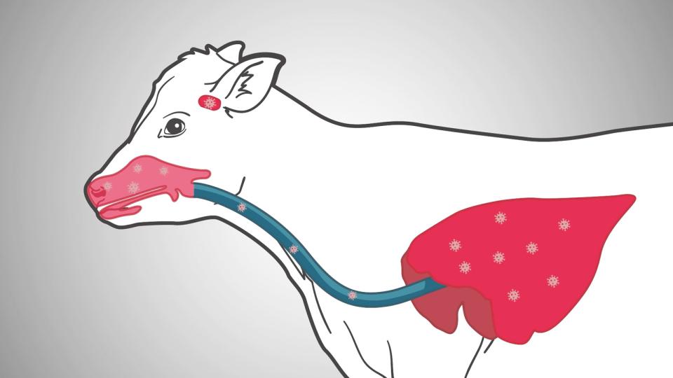
Taking the respiratory syndrome first, IBR typically involves high morbidity (80-100%) but low mortality (<2%). Affected animals present with a pyrexia often in excess of 40oC, increased respiration rate, depression and lethargy, anorexia and the characteristic serous nasal discharge. White plaques may be visible in the nasal mucosa and on the tongue and harsh respiratory sounds are audible over the trachea due to the necrosis of the mucosa.

The inflammation and necrosis of the nasal and tracheal mucosa often leads to secondary bacterial involvement, initially in the upper airway, leading to the nasal discharge turning mucopurulent (usually after 72hrs or so) and can go on to cause a bacterial bronchopneumonia (usually within 7-10 days of onset of clinical signs).

Many (but not all) cases will also present with the conjunctival form with initially serous, progressing to purulent, discharge from either one or both eyes. There may also be white plaques (caused by aggregations of lymphocytes) on the palpebral conjunctiva.

Co-infection with M. haemolytica in the lungs and Moraxella bovis in the conjunctival presentation can have synergistic effects in the animals, increasing the pathogenicity of the infection. In the case of secondary pneumonic infection with M. haemolytica, mortality rates can increase significantly.  

In adult animals this will be accompanied by severe milk drop which may (especially in heifers) result in depressed yield for the remainder of the lactation. Abortions are also common, caused by both the pyrexic response and direct foetal infection. Abortions can occur during the acute phase of the infection or up to 2 months later.

The encephalitic form is considerably rarer and generally presents in calves. It is often accompanied by focal white plaques on the tongue. Animals may present with a range of neurological signs from blindness and recumbency to opisthotonos and convulsions.

Pustular vulvovaginitis or infectious balanoposthitis are venereal presentations of BoHV-1 infection. They can be independent of the respiratory presentations or coincidental. The condition can be spread venereally and results in ulcerative white plaques and erosions on the vulval and vaginal mucosa, which may be uncomfortable for the cow on palpation. These plaques often occur fairly soon after intercourse with another infected animal, often with 2-3 days. Abortion is seen albeit less commonly than in the respiratory presentations.

Pathophysiology

In the rhinotracheitis presentation, BoHV-1 enters the mucosal cells and replicates causing widespread cell death and inflammation. This is usually confined to nasal cavity, para-nasal sinuses and trachea all of which become severely oedematous and produce prodigious amounts of serous, then mucous discharge. The inflammation and oedema in the trachea may be so severe that the wall swells to 2cm thick causing severe stenosis of the trachea and producing the characteristic rales on auscultation.

In these initial stages of the clinical disease, the immune response is predominantly cell mediated with significant neutrophil and mononuclear infiltration into the affected tissues. This inflammation affects both the mucosa and the submucosa leading to intense ulceration and necrosis of the mucosa of the nasopharynx and trachea along with significant fibrin deposition. It is this sloughing of necrotic and fibrinous epithelial tissue which gives rise to the oft seen secondary bacterial pneumonia. This secondary bacterial pneumonia is also enabled further by IBR infection by way of a concurrent reduction in T-cell population, possibly by penetrating CD4+ T cells and initiating apoptosis.

It is during this acute phase of infection with IBR that the BoHV1 penetrates the ganglionic neurons (usually trigeminal and the sensory neurons) and, while the acute infection may be cleared, the latency-associated transcript of the viral gene expression persists in the ganglia, often for the rest of the cow’s life. This may then be reactivated by stress events or immunosuppression; indeed recrudescence has been demonstrated with administration of dexamethasone 10weeks post infection. This then leads to viral shedding and, frequently, reappearance of the clinical syndrome. The immune response for these subsequent bouts of disease is usually an antibody response.

The antibody response is believed to be seroconversion for life however, the efficacy of the antibody response does wane and will be notably poorer 6 months later, after which time the animal will be susceptible to clinical disease again from the recrudescence of the latent virus in their own trigeminal nerve ganglia.

IBR virus

Epidemiology

IBR is prevalent in most countries worldwide with the exception of a few countries in Europe. The Scandinavian nations (Sweden, Norway, Denmark and Finland), Switzerland, the Czech Republic, Austria and Germany along with some Channel islands in the UK are officially free of IBR. Belgium, Italy, Spain and the Netherlands have national or regional eradication schemes.

In order to be considered IBR free, 99.8% of animals nationally have to be on IBR negative holdings. As a comparator the current prevalence of herds testing positive for IBR in the UK is between 43-84%.

Current published data for prevalence in Brazil suggests, within the Caparao region, 48.59% of cattle are seropositive with 100% of herds tested containing seropositive animals, and in the state of Parana 41.9% of animals were seropositive with 90.5% of herds containing seropositive animals. Experience in other nations tells us that, without structured intervention and robust vaccination policies using 6 monthly immunisations, this prevalence is likely to increase and will certainly be causing significant production losses.

Diagnosis

Initial diagnosis of the respiratory or venereal conditions can be made on the basis of clinical signs.

On necropsy examination, the tracheal mucosa will be red and thickened and covered in mucopurulent exudate. The oedema can be significant enough to dramatically reduce the diameter of the lumen of the trachea which results in the tracheal stertor heard in the acute phase on auscultation. The mucosal surfaces of the nasopharynx will also be severely congested and covered in mucopurulent discharge and may also show areas of petechial haemorrhage.

In the acute stage of the clinical disease Fluorescent Antibody tests (FAT) can be run on swabs from the nasopharynx, conjunctiva or vagina (depending on the presentation) as can PCR tests which can also be run on tissue samples including lung, lymph node, kidney, spleen or thymus however this test is not approved for use in the UK.

Paired serology can also be used to test ongoing outbreaks albeit with a delay to a confirmatory result in comparison to the FAT test.

Modern vaccines have the glycoprotein E antigen deleted allowing for a DIVA test to confirm “wild type” vs vaccinated status. It is still possible to obtain non-marker vaccines in the UK in multivalent calf vaccines and as such, a thorough history of vaccinations on the unit must be taken to assess whether this would interfere with testing. The antibodies are sufficiently long lived to interfere with bulk milk monitoring and their use on farms now should be seriously questioned.

Vaccinations

The first IBR vaccines were developed with the aim of preventing clinical signs of disease (i.e. traditional multivalent vaccines). More recently, marker vaccines were introduced using the DIVA concept (differentiating infected from vaccinated animals). The use of marker vaccines together with a diagnostic test capable of distinguishing which animals have been vaccinated or naturally infected is key to control (or even eradicate) IBR. Marker vaccines lack a glycoprotein (gE deletion) while they can elicit an immune response against the rest of antigens (i.e. gB); a serology test (gE blocking ELISA) can detect which animals have been vaccinated (gE negative/gB positive) or infected (gE positive/gB positive).

A more modern marker vaccine is available in Europe and the Americas, it contains not only a gE deletion but a second deletion in the thymidine kinase (tk) gene, which is associated to viral neurotropism and latency. This extra deletion increases safety: it decreases the likelihood of producing latency and further reduces the virulence of the vaccine strain.

Marker vaccines allow implementing strategies to gradually reduce seroprevalence. The use of marker vaccines every six months (hyperimmunization) has proven to be effective to control and eradicate IBR in some European countries (i.e. Germany, Czech Rep.). Likewise, hyperimmunization has been successfully used to control and eradicate Alphaherpesvirus in other species such as Aujeszki in swine. Actually, several studies have demonstrated how hyperimmunization with inactivated and, to a greater degree, live marker vaccines reduced the reproduction ratio, proving the prevention in virus circulation in vaccinated herds. The reduction of IBR (gE) seroprevalence by hyperimmunization with inactivated and live attenuated vaccines has also been demonstrated in a longitudinal field trial. This, six-monthly vaccination has proven its efficacy in reducing virus circulation when the whole herd is vaccinated.

Conclusion

IBR remains a significant contributor to bovine respiratory disease complex and has significant effects on production and fertility in cattle herds. Its classical respiratory syndrome with involvement of the conjunctiva and the upper respiratory tract remains instantly recognisable and the highly infectious nature of the virus makes control in the face of an outbreak challenging. The typical herpesvirus facet of latency has made its eradication extremely difficult in most countries with only a handful of European nations making headway with eradication programs however control is possible with robust, 6 monthly vaccination protocols and monitoring.


Author: Oliver Maxwell BVSc BSc(Hons) MVM DipECBHM. Royal College Recognised Specialist in Cattle Health and Production. European Specialist in Bovine Health Management


References:

Ampe B., Duchateau L., Speybroeck N., Berkvens D., Dupont A., Kerkhofs P., Thiry E., Dispas M., 2012.Assessment of the long-term effect of vaccination on transmission of infectious bovine rhinotracheitis virus in cattle herds hyperimmunized with glycoprotein E–deleted marker vaccine  American journal of veterinary research, 73(11), pp.1787-1793.

Brar, J.S., Johnson, D.W., Muscoplat, C.C., Shope Jr, R.E. and Meiske, J.C., 1978. Maternal immunity to infectious bovine rhinotracheitis and bovine viral diarrhea viruses: duration and effect on vaccination in young calves. American journal of veterinary research, 39(2), pp.241-244.

Bosch J.C., De Jong M.C.M., Franken P., Frankenas K., Hage J.J., Kaashoek M.J., Maris-Veldhuis M.A., Noordhuizen J.P.T.M., Van der PoeI W.H.M., Verhoeff J., Weerdmeester K., Zimmer G.M., Van Oirschot J.T., 1998 An inactivated gE negative marker vaccine and an experimental gD-subun:it vaccine reduce the incidence of bovine herpesvirus 1infections in the field. Vaccine, 16, pp.265-271.

Carter, J.J., Weinberg, A.D., Pollard, A., Reeves, R., Magnuson, J.A. and Magnuson, N.S., 1989. Inhibition of T-lymphocyte mitogenic responses and effects on cell functions by bovine herpesvirus 1. Journal of Virology, 63(4), pp.1525-1530.

D’arce, R.C.F., Almeida, R.S., Silva, T.C., Franco, A.C., Spilki, F., Roehe, P.M. and Arns, C.W., 2002. Restriction endonuclease and monoclonal antibody analysis of Brazilian isolates of bovine herpesviruses types 1 and 5. Veterinary microbiology, 88(4), pp.315-324.

Davies, D.H. and Carmichael, L.E., 1973. Role of cell-mediated immunity in the recovery of cattle from primary and recurrent infections with infectious bovine rhinotracheitis virus. Infection and Immunity, 8(4), pp.510-518.

Griebel, P.J., Ohmann, H.B., Lawman, M.J.P. and Babiuk, L.A., 1990. The interaction between bovine herpesvirus type 1 and activated bovine T lymphocytes. Journal of General Virology, 71(2), pp.369-377.

Griebel, P.J., Qualtiere, L., Davis, W.C., Lawman, M.J. and Babiul, L.A., 1987. Bovine peripheral blood leukocyte subpopulation dynamics following a primary bovine herpesvirus-1 infection. Viral immunology, 1(4), pp.267-286.

Jones, C., 2003. Herpes simplex virus type 1 and bovine herpesvirus 1 latency. Clinical microbiology reviews, 16(1), pp.79-95.

Mars M.H., de Jong M.C.M., Franken P., van Oirschot J.T., 2001.Efficacy of a live glycoprotein E-negative bovine herpesvirus 1 vaccine in cattle in the field. Vaccine, 19 (15-16), pp.1924-1930.

Muylkens, B., Thiry, J., Kirten, P., Schynts, F. and Thiry, E., 2007. Bovine herpesvirus 1 infection and infectious bovine rhinotracheitis. Veterinary research, 38(2), pp.181-209.

Pastoret, P.P., Aguilar-Setien, A., Burtonboy, G., Mager, J., Jetteur, P. and Schoenaers, F., 1979. The effect of repeated treatment with dexamethasone on the re-excretion pattern of infectious bovine rhinotracheitis virus and humoral immune response. Veterinary Microbiology, 4(2), pp.149-155.

Petrini S, Iscaro C, Righi C. Antibody Responses to Bovine Alphaherpesvirus 1 (BoHV-1) in Passively Immunized Calves., 2019. Viruses, 11(1) 23.

Spilki, F.R., Esteves, P.A., Lima, M.D., Franco, A.C., Chiminazzo, C., Flores, E.F., Weiblen, R., Driemeier, D. and Roehe, P.M., 2004. Comparative pathogenicity of bovine herpesvirus 1 (BHV-1) subtypes 1 (BHV-1.1) and 2a (BHV-1.2 a). Pesquisa Veterinária Brasileira, 24(1), pp.43-49.

Stabel, J.R., Spears, J.W. and Brown Jr, T.T., 1993. Effect of copper deficiency on tissue, blood characteristics, and immune function of calves challenged with infectious bovine rhinotracheitis virus and Pasteurella hemolytica. Journal of animal science, 71(5), pp.1247-1255.

Vannie, P., Capua, I., Le Potier, M. F., Mackay, D. K., Muylkens, B., Parida, S., Paton, D. J., & Thiry, E., 2007. Marker vaccines and the impact of their use on diagnosis and prophylactic measures. Revue scientifique et technique (International Office of Epizootics), 26(2), 351–372.

Wentink, G.H., Van Oirschot, J.T. and Verhoeff, J., 1993. Risk of infection with bovine herpes virus 1 (BHV1): a review. Veterinary Quarterly, 15(1), pp.30-33.

Winkler, M.T.C., Doster, A. and Jones, C., 1999. Bovine herpesvirus 1 can infect CD4+ T lymphocytes and induce programmed cell death during acute infection of cattle. Journal of virology, 73(10), pp.8657-8668.